
Fig. 3.
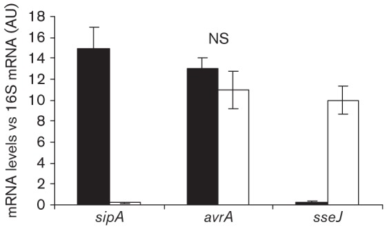
Analysis of avrA expression in HEp-2 cells by qRT-PCR. Epithelial cells were infected with a wt strain of S. Enteritidis for 20 min (black bars) or 10 h (white bars). Post-infection cells were processed as indicated in Methods to obtain total mRNA. sipA, avrA and sseJ mRNA levels from bacteria colonizing HEp-2 cells were measured by qRT-PCR at the indicated times. sipA and sseJ were used as controls for SPI-1 and SPI-2 expression, respectively. The mRNA amount was related to 16S mRNA levels. Values are means±sd of three independent mRNA extractions performed in triplicate. AU, Arbitrary unit; NS, no significant difference between the two times post-infection (ANOVA).