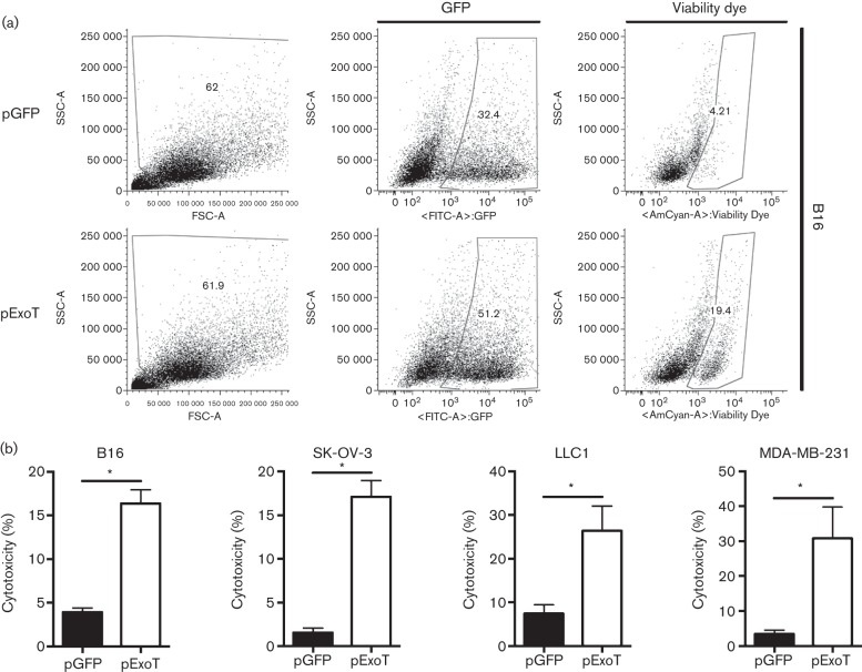
Fig. 4.
Assessment of ExoT-induced cytotoxicity in cancer by Viability Dye. Cancer cells were transfected with pGFP vector control or pExoT-GFP. At 17 h after transfection, cells were stained with Viability Dye and analysed by flow cytometry. (a) Representative FACS plots for B16 show ExoT was sufficient to cause cytotoxicity in transfected cells. SSC, side scatter; FSC, forward scatter. (b) FACS analysis of transfected B16 cells indicated that pExoT-GFP resulted in significantly more cytotoxicity compared with pGFP-transfected cells. *P<0.0001; n = 7; Student’s two-tailed t-test.

