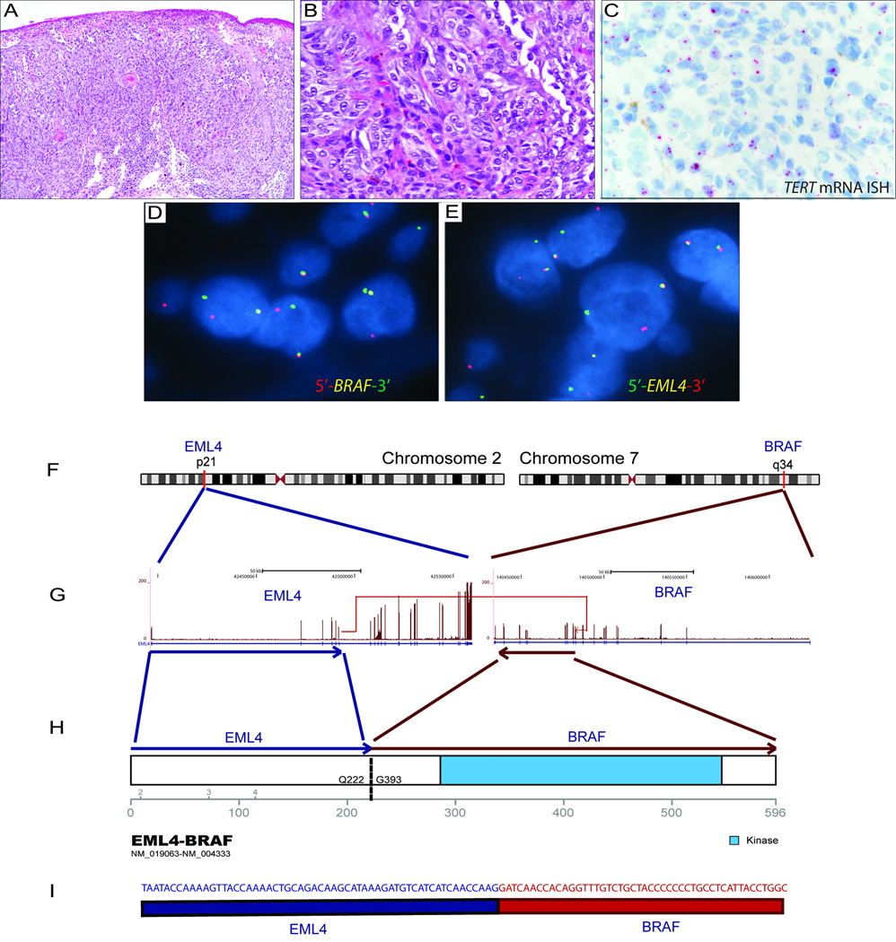
Figure 1. Spitzoid melanoma with the EML4–BRAF fusion transcript.
Analysis of a tumor sample from a 14 year-old male (patient 1) with a 1.3-mm-thick spitzoid melanoma with microscopic sentinel lymph node metastasis at diagnosis and death as a result of disseminated disease in 18 months. (A, B) H&E photomicrographs (10× and 40×) of the primary tumor show compound proliferation of epithelioid and spindle-shaped spitzoid melanocytes arranged in confluent junctional nests with ulceration. (C) TERT mRNA ISH shows bright intracellular signals in melanocytes in this TERT promoter mutant melanoma. (D, E) Break-apart FISH shows splits of the red and green signals, which is consistent with the rearrangement of BRAF and EML4, respectively. (F) Schematic figure depicting the reciprocal translocation between 2p21 and 7q34, resulting in the chimeric products EML4–BRAF and BRAF–EML4. (G) The in-frame fusion transcript produced by joining exon 6 of EML4 (NM_019063, chr2:42491872) to exon 10 of BRAF (NM_004333, chr7:140482957). (H) The fusion transcript results in a 596-amino-acid chimeric protein containing the intact kinase domain of BRAF. (I) The RNA transcript contig shows the fusion breakpoint in the EML4–BRAF chimeric gene.

