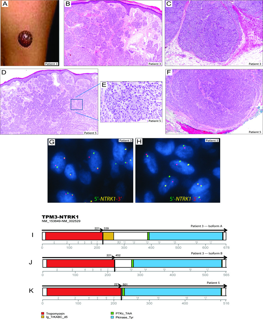
Figure 3. Multiple isoforms of the TPM3–NTRK1 fusion transcripts in 2 spitzoid melanocytic tumors.
(A) Photograph of a 2-year-old boy (patient 3) with an amelanotic exophytic nodule on the thigh, which was clinically thought to be a pyogenic granuloma. (B) H&E photomicrograph (4×) of the upper part of the lesion shows fascicles of spindled and epithelioid melanocytes arranged in whorls and nests with edema of the papillary dermis with telangiectasia. There were lymphovascular invasion and focal necrosis (not shown). (C) H&E photomicrograph (4×) of the bottom part of the lesion shows dumbbell-shaped configuration and deep extension into the subcutaneous fat. (D and F) H&E photomicrographs (4×) of the top and bottom portions of a lesion on the ear of a 6-year-old female (patient 5) showing nests of polygonal shaped epithelioid melanocytes at high magnification (inset in E) that extend throughout the dermis and far into the basal line of resection margin in subcutis. (G, H) Break-apart FISH for NTRK1 shows split signals in both spitzoid tumors. (I–K) The NTRK1 fusion gene encoded 2 variant TPM3–NTRK1 isoforms in patient 3 and a third isoform in patient 5. All isoforms retained the intact tyrosine kinase domain of NTRK1.

