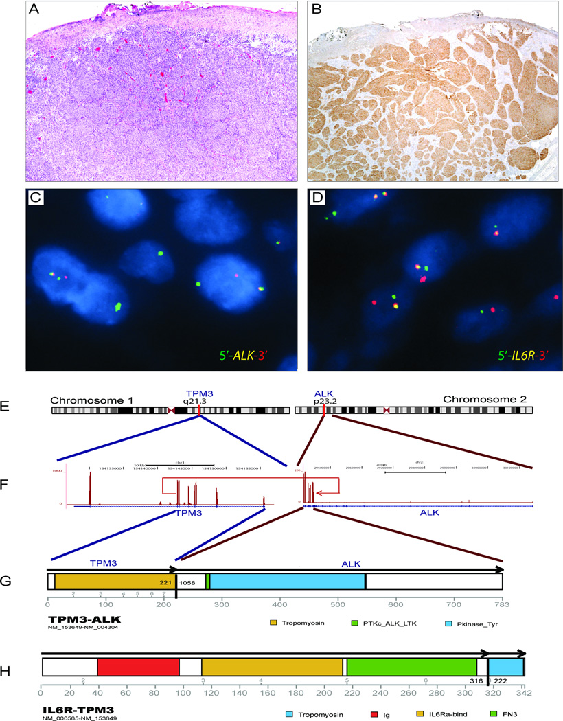
Figure 4. Complex genomic rearrangements involving ALK, TPM3, and IL6R.
Spitzoid melanocytic neoplasm in the calf of a 13-year-old female (patient 4). (A) H&E photomicrograph showing nests of spindled melanocytes with ulceration. (B) ALK immunoreactivity highlights a fascicular architecture in the dermis. (C, D) Break-apart FISH for ALK and IL6R confirms rearrangements in the respective genes. (E–H) Schematic of the TPM3–ALK fusion gene. The first 222 amino acids of TPM3 are fused to the C terminus of ALK, incorporating the intact tyrosine kinase domain of ALK. The remaining TPM3 protein is fused to the IL6R protein. The identification of multiple fusions involving TPM3 suggests a complex translocation event.

