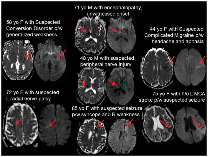
Figure 2. Hyperacute MRI diffusion weighted imaging for patients receiving IV tPA.

Brief clinical histories and diffusion weighted imaging (DWI) are shown for the 7 patients who were screened with hMRI to aid tPA-decision making and who received IV tPA. Clinical histories are consistent with suspected stroke mimics suggesting likely appropriate utilization of hMRI. Acute ischemic stroke lesions (red arrows) shown as hypointensity on apparent diffusion coefficient (ADC) maps (left) and hyperintensity on diffusion weighted imaging (right) for each patient demonstrate small lesion areas that appear to be of recent onset (given mild hyperintensity on DWI).
