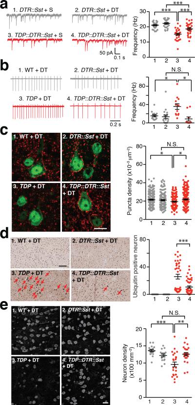
Figure 3. Bilateral ablation of Sst interneurons diminished excitotoxicity of L5-PN in TDP mice.
(a) Two-week bilateral ablation of Sst interneurons in M1 cortex of TDP mice increased L5-PN mIPSC frequency. Left: representative traces of L5-PN mIPSCs. Right: dot plots of the frequencies of mIPSCs (S, saline; Group 1 to 4, n = 25, 26, 27, 28 neurons, 3 mice; One-way ANOVA and post hoc Tukey test). (b) Two-week bilateral ablation of Sst interneurons in M1 restored normal excitability of L5-PN in TDP mice. Left: representative traces of AP firing of L5-PN, through loose-seal cell-attached recordings with bath-application of 20 mM KCl. Right: dot plots of AP firing frequencies (Group 1 to 4, n = 15, 18, 12, 10 neurons, 3, 4, 4, 3 mice; Brown and Forsythe Test and post hoc Games-Howell test). (c) Six-week bilateral ablation of Sst interneurons in M1 cortex of TDP mice increased vesicular GABA transporter (VGAT) punta in L5-PN. Left: representative images of VGAT staining (Red, VGAT; Green, NeuN; scale bar, 10 μm). Right: dot plots of puncta densities (Group 1 to 4, n = 180, 120, 143, 195 neurons, 3, 2, 3, 3 mice; Brown and Forsythe test and post hoc Games-Howell test). (d) Six-week bilateral ablation of Sst interneurons reduced ubiquitin positive aggregates in TDP mice. Left: representative images (red arrows indicated ubiquitin positive neurons; scale bar, 100 μm). Right: dot plots of ubiquitin positive neuron numbers (Group 1 to 4, n = 24, 24, 36, 42 counts, from 12, 12, 18, 21 slices of 4, 4, 6, 7 mice; Mann-Whitney U-test). (e) Six-week bilateral ablation of Sst interneurons reversed neuronal loss in M1 cortex of TDP mice. Left: representative images of NeuN immunostaining (scale bar, 20 μm). Right: dot plots of layer 5 neuron densities (Group 1 to 4, n = 18, 16, 24, 24 counts from 9, 8, 12, 12 slices of 3, 3, 4, 4 mice; Brown and Forsythe test and post hoc Games-Howell test). Error bars are mean ± SEM. *, p < 0.05; **, p < 0.01; ***, p < 0.001; N.S., not significant.

