
Figure 2. MYXV indices apoptosis in primary MM cells.
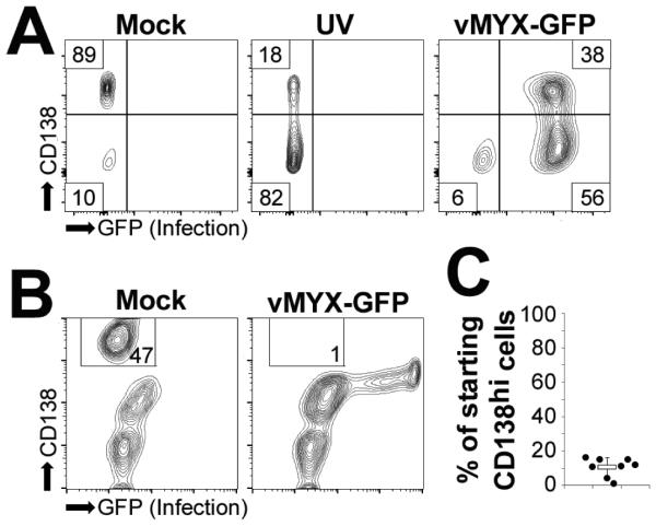
(A) Human U266 MM cells were either mock-treated, subjected to UV light for one hour, or infected with vMYX-GFP at an MOI=10. Six hours after treatment, cell surface expression of CD138 was analyzed using flowcytometry. (B) Human bone marrow aspirates depleted of red blood cells were either mock-treated or infected with vMYX-GFP at an MOI=10. Six hours post-infection cell surface expression of CD138 was analyzed using flowcytometry. (C) Quantitation of ‘B’. Data presented as percent of starting CD138hi cells which was calculated by comparing the number of CD138hi cells in mock and vMYX-GFP infected samples.