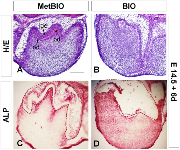
Figure 2.

E14.5 first molars cultured with MetBIO (control) or BIO (20 μM) for 6 days. Control molar sections stained with hematoxylin-eosin (A) had normal development of dental cusps, clear polarization of the inner dental epithelia cells (ide), odontoblast differentiation (od), and a thin layer of predentin (pd). In contrast, molars cultured with BIO treatment (B) showed an irregular and disorganized dental cusp pattern, non-polarized cells in the inner dental epithelium and an undifferentiated odontoblastic layer. Alkaline phosphatase activity spread all over the dental mesenchyme in BIO-treated molars (D), but in control samples, enzymatic activity was detected in only in the odontoblastic and subodontoblastic layers (C). The dotted region corresponds to dental epithelium. Od, odontoblasts; ide, inner dental epithelium; pd, predentin. Scale bar: 200 μm.
