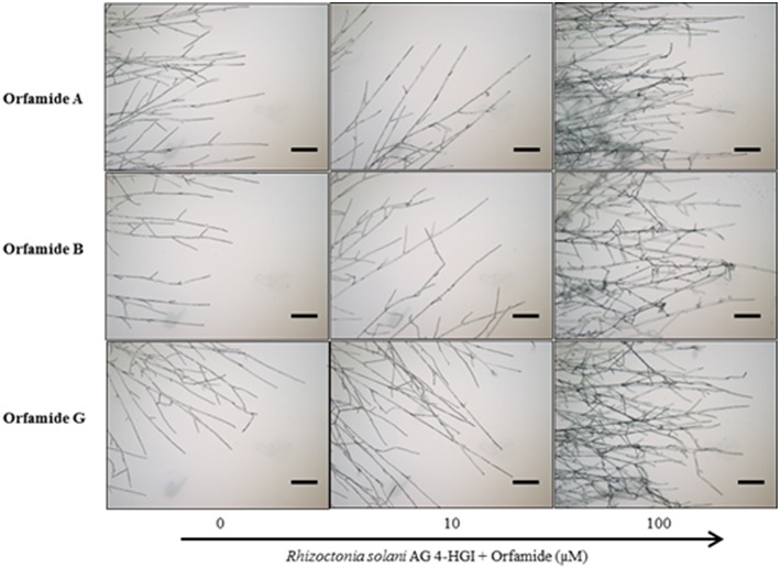
Figure 5.
Microscopic assays showing the effect of various concentrations of orfamides (Orfamide A, orfamide B, and orfamide G) on hyphal branching of Rhizoctonia solani AG 4-HGI, scale bar = 100 μm. Sterile microscopic glass slides were covered with a thin, flat layer of water agar (Bacto agar; Difco) and placed in a plastic Petri dish containing moist sterile filter paper. An agar plug (Diameter = 5 mm) taken from an actively growing colony of R. solani was inoculated at the center of each glass slide. Two droplets (15 μl each) containing 0, 10, or 100 μM orfamide were placed at two sides of the glass slide at about 2 cm from the fungal plug. Slides were incubated for 36 h at 28°C before evaluation under an Olympus BX51 microscope.

