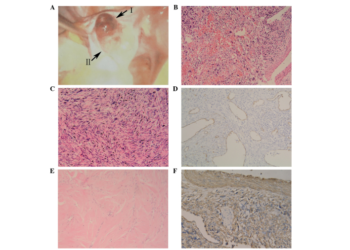
Figure 1.
Imaging studies of the ovarian mass. (A) Exploratory laparoscopy revealing a mass in the enlarged left ovary, composed of two regions (regions I and II). Microscopic examination of region I revealed (B) focal hemorrhage and necrosis (hematoxylin and eosin staining; magnification, x100) (C) with pleomorphic and fusiform cells (hematoxylin and eosin staining; magnification, x200). (D) Blood vessels demonstrating an absence of adventitial layers (blood vessel wall exhibiting intense immunostaining for cluster of differentiation 34; magnification, x100) in a section of region I. (E) Microscopic examination of region II (hematoxylin and eosin staining; magnification, x100). (F) Immunohistochemical staining of region I was SMA+ (ovarian leiomyosarcoma demonstrating intense immunostaining for SMA; magnification, x200). SMA, smooth muscle actin.

