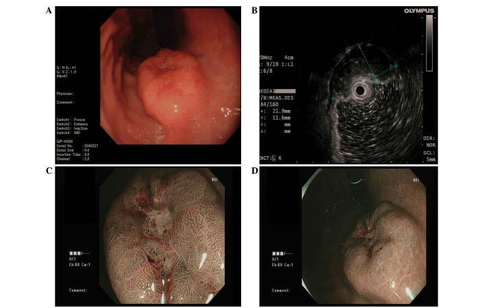
Figure 1.
(A) Gastroscopy revealed a submucosal tumor-like mass covering the anterior wall of the gastric body, with a surface ulcer on top of the lesion. (B) Endoscopic ultrasound demonstrated a hypoechoic lesion originating from the submucosal layer. (C and D) Narrow band imaging of the tumor suggested no marked glandular ducts in the central area, and normal glandular ducts in the peripheral area.

