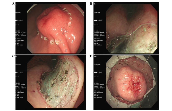
Figure 3.
ESD was performed for definitive diagnosis and treatment of the gastric lesion. (A) The perimeter of the lesion was marked with a cautery knife. (B) The submucosa beneath the lesion was injected and dissected using a surgical knife. (C) The specimen in the stomach was completely resected. (D) ESD of the lesion (2.5×2.5×0.4 cm) was successful and the specimens were obtained after obtaining consent from the patient. ESD, endoscopic submucosal dissection.

