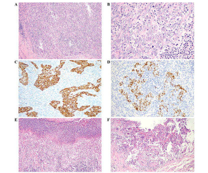
Figure 4.
Pathology of ESD specimen of the gastric lesion. The histopathological and immunohistochemical examinations were compatible with LELGC. The basal margin was positive for carcinoma whereas the lateral margin was negative. The tumor had invaded the submucosal layer. (A and B) The tumor infiltrated uniformly with an abundance of lymphocytes and plasma cells throughout the entire area of the tumor. [Hematoxylin and eosin staining; (A) magnification, x100, (B) magnification, x400]. (C) Cytokeratin expression was positive in the LELGC (Envision double staining; magnification, x200) and (D) Epstein-Barr virus-encoded RNA in situ hybridization also was positive (magnification, x200). Postoperatively, lymphoepithelioma-like carcinoma of the stomach was diagnosed and staged as IA T1bN0cM0 according to the Tumor-Node-Metastasis classification of gastric carcinoma. (E) Ulceration was observed in the ESD specimen (hematoxylin and eosin staining; magnification, x100); (F) no carcinoma tissue was found in the ulcer (hematoxylin and eosin staining; magnification, x200). ESD, endoscopic submucosal dissection; LELGC, lymphoepithelioma-like gastric carcinoma.

