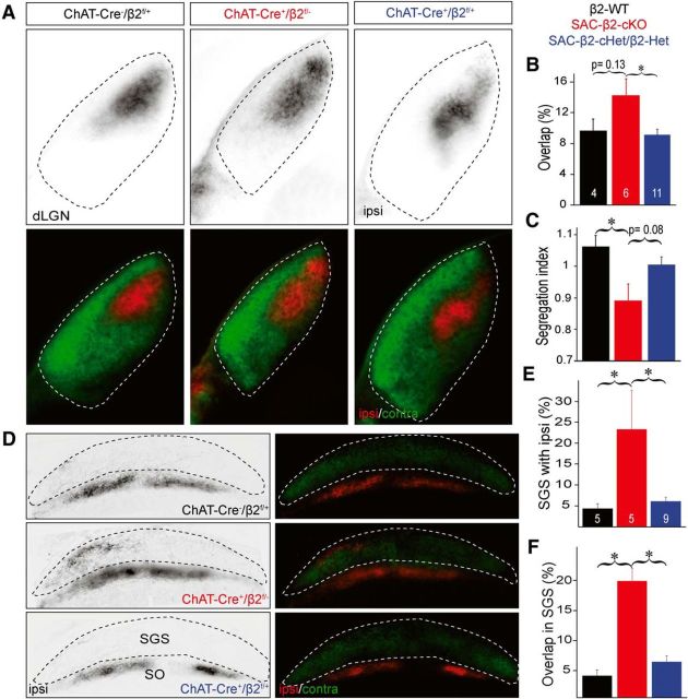
Figure 5.
Eye-specific segregation is disrupted in the SAC-β2-cKOs at P8. Whole eye (vitreal) injection of Alexa-conjugated cholera toxin dye bulk labels most RGC axon projections in the dLGN (A–C) and the SC (D–F). A, Example dLGN (coronal sections) of WT (ChAT-Cre−/β2fl/+) and β2 heterozygous controls (ChAT-Cre+/β2fl/+; SAC-β2-cHet and ChAT-Cre−/β2fl/−; Het; combined in B, C, E, F). RGC projections from the contralateral eye (green) and the ipsilateral eye (red). B, Summary histogram showing the percentage of dLGN area with overlapped contralateral and ipsilateral projections. In SAC-β2-cKOs (ChAT-Cre+/β2fl/fl), ipsilateral eye projections were expanded and intermingled with projections from the contralateral eye, resulting in a greater overlap relative to controls. C, Summary histogram showing segregation of contralateral and ipsilateral projections (Segregation index) in dLGN. Segregation index was reduced in SAC-β2-cKOs relative to controls (SAC-β2-cHet and Het controls combined). D, Example retinocollicular projections from WT, β2 heterozygous controls, and SAC-β2-cKO mice. Contralateral axons (green) project to the most superficial (SGS) layer of the SC (sagittal sections), and ipsilateral axons (red) project to a layer (SO) inferior to the contralateral axons. E, Summary quantification of the percentage of the SGS layer with ipsilateral projections. In SAC-β2-cKOs, more ipsilateral eye axons extended into the SGS layer than in control mice. F, Summary quantification of percentage of SGS layer with projections that overlap between the ipsilateral and contralateral eye. In SAC-β2-cKOs, ipsilateral eye axons overlapped to a greater extent with contralateral axons than in control mice, indicating poor eye-specific segregation. R, Rostral; C, caudal; SO, stratum opticum. *p < 0.05 (ANOVA with Tukey's HSD). Error bars indicate SEM. Scale bars, 500 μm.

