Figure 7.
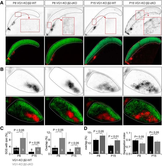
Eye-specific projections from retina to SC were disrupted in VG1-KO/β2-cKOs at P8 and P15. A, Examples of eye-specific projections to the SC in mice with combined manipulations of Vglut1 and β2 expression at P8 (left) and P15 (right). Top, RGC axon projections from the ipsilateral eye to the SC. Bottom, RGC projections from both eyes to the SC. In VG1-KO/β2-WT controls (VG1−/−/ChAT-Cre−/β2fl/+), axons from the contralateral eye projected to the superficial layer (SGS) of the SC and axons from the ipsilateral eye projected to the stratum opticum. Genetic deletion of Vglut1 alone had no effect on eye-specific projections at P8 (Fig. 6). Additional conditional deletion of β2 from SACs disrupted the eye-specific projections at P8 and P15. RGC axon arbors from the ipsilateral eye projected to the SGS layer and overlapped with projections from the contralateral eye in DKO mice. B, Examples of eye-specific projections to the dLGN in the same groups of mice at P8 and P15. Top, RGC axon projections from the ipsilateral eye to the dLGN. Bottom, RGC projections from both eyes to the dLGN. Disrupted binocular projections in SAC-β2-cKO mice at P8 (Fig. 5) were not restored at P15 in mice that also lack VG1 (DKO). C, Summary quantification of eye-specific segregation in the SC. At P8, deletion of VG1 and conditional deletion of β2 from SACs significantly increased ipsilateral arbors in the SGS layer. Ipsilateral projections were still disrupted in DKOs at P15 relative to controls. D, Summary quantification of eye-specific segregation in the dLGN. Segregation was worse in mice that lack both VG1 and β2 (DKO) in SACs at P8 and P15.

