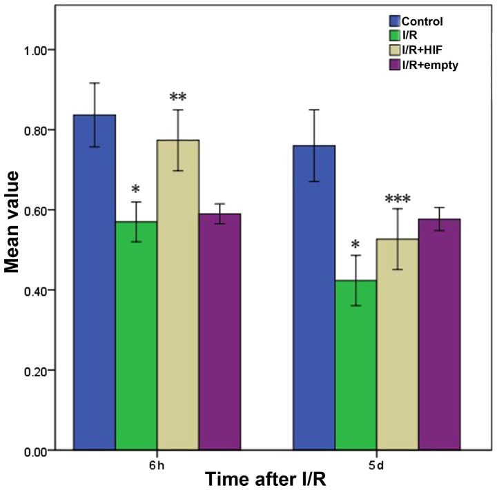
Figure 2.
Myocardial cell viability based on the methyl thiazolyl tetrazolium assay at 6 h or 5 days following treatments simulating I/R. Control cells were subjected to mock treatment; I/R cells were subjected to I/R; HIF-1α cells were transiently transfected with a plasmid expressing full-length HIF-1α prior to I/R; and empty cells were transiently transfected with empty expression vector prior to I/R. The data are shown as the mean ± standard deviation for three independent experiments. *P<0.05, vs. control; **P<0.05, vs. untransfected cells; ***P<0.05, vs. 6 h after I/R or mock treatment. I/R, ischemia-reperfusion; HIF-1α, hypoxia-inducible factor-1α.

