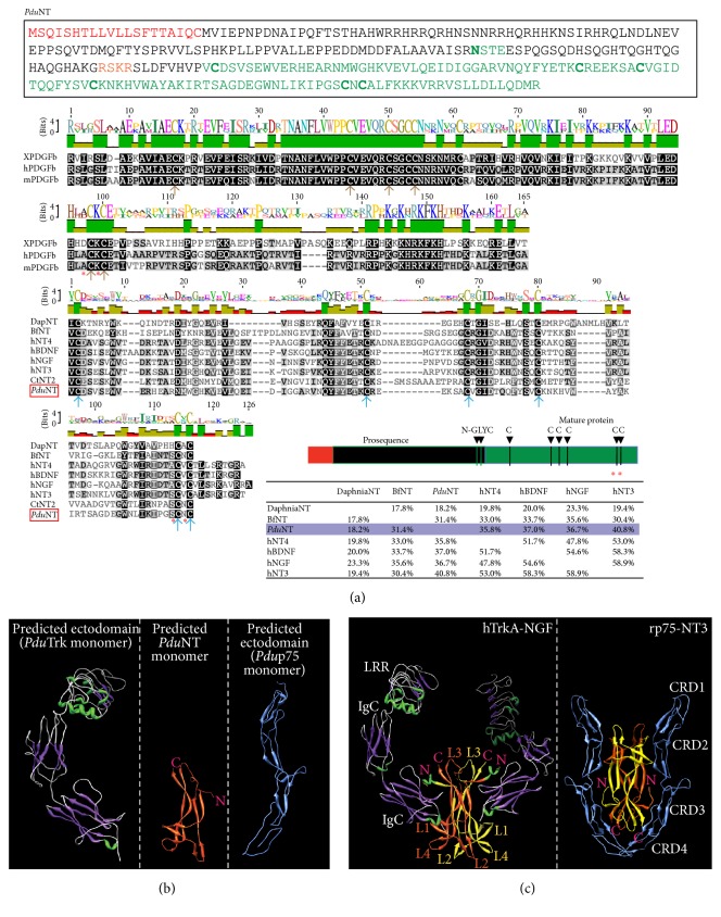
Figure 2.
Sequence analysis of PduNT and 3D modeling of the extracellular domain of PduTrk, NT, and p75. (a) Predicted a.a. sequence, schematic representation, and multiple sequence alignment for Platynereis NT. The alignment is done for the “Cys-Knot” domain (mature protein). In comparison the Cys-Knot of PDGF-beta is also shown. In the schematics in the lower right the different domains and the cysteines core (green) are indicated. Red labels the predicted signal peptide. N-glyc: putative glycosylation site. Species are indicated as in Figure 1. In the alignment, the cysteines forming the knot are shown with arrows (brown for the PDGF subfamily, blue for the NGF subfamily). (b) Predicted 3D structure of the extracellular domain of PduTrk (left panel), PduNT (middle panel), and Pdup75 (right panel). (c) As reference, a published 3D structure of the complex between the extracellular domain of TrkA-NGF (left panel, DOI: 10.2210/pdb1www/pdb [23]) and p75-NT3 (right panel, DOI: 10.2210/pdb3buk/pdb [24]) is shown. The domains are indicated (see text for details), as well as the N-terminus (N) and C-terminus (C) of the NTs (in pink), and the 4 loops formed by the NGF dimmer.

