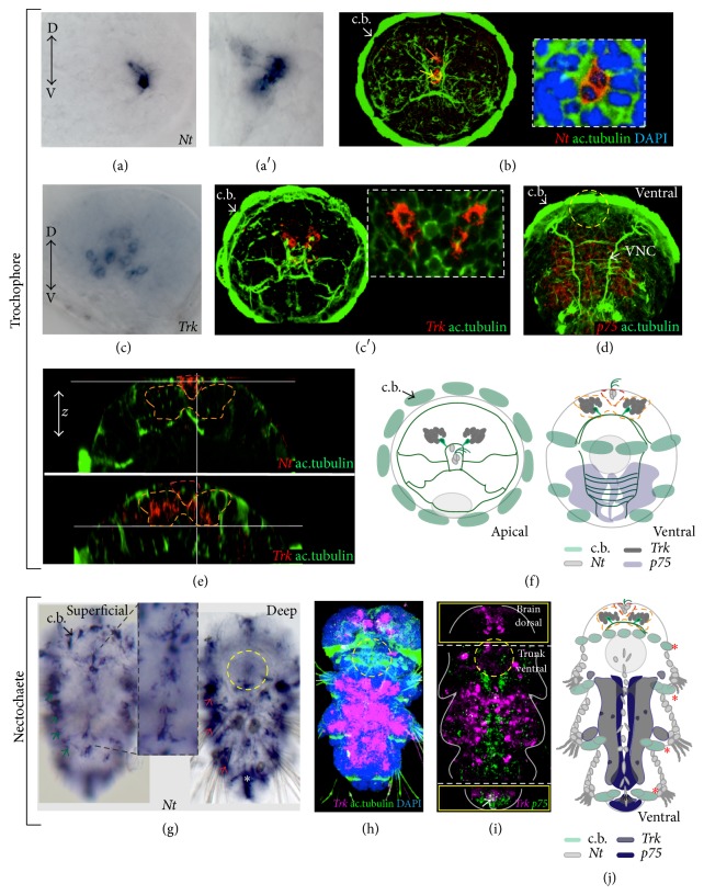
Figure 4.
Expression of PduNT, p75, and Trk in the developing worm. (a–b) Apical views. Expression of Nt in cells of the apical organ at 48 hpf ((a′) and the inset in (b) show a close-up on the Nt+ cells). Bright field images (a, a′), confocal z-projections (b). The yellow arrow indicates the apical tuft cells; the orange arrow indicates the crescent cells. (c, c′) Apical views. Expression of Trk in cells in the dorsal brain (the inset in (c′) shows a close-up on the Trk+ cells). Bright field image (c), confocal z-projection (c′). (d) Ventral view. Expression of p75 in the developing nervous system of the trunk, VNC: ventral nerve cord, confocal z-projection. In (a)–(d), D: dorsal, V: ventral. (e) Virtual cross sections show the position of the Nt+ (dashed red contour)/Trk+ cells (dashed orange contour) along the z-axis. (f) Schematic drawings of the apical view (left) and ventral view (right) showing Nt (light gray), Trk (dark gray), and p75 (light blue) expression of the trochophore larvae. (g) Superficial and deep Nt expression in the juvenile larva (nectochaete, around 3 dpf). Expression in the ciliary band (c.b.) along the superficial (green arrows) and deep (red arrows) periphery and in the midline (inset in (g)) is observed. (h) Trk expression in the ventral trunk nervous system and in the brain in the nectochaete stage. (i) Double WMISH showing the expression of Trk and p75 in the juvenile larva. The white arrow indicates colocalization at the posterior end. Different Z-projections from the same confocal scan are outlined by yellow squares: the upper one shows a single channel (magenta for Trk) within a z-projection of the dorsal brain (p75 mRNA is not found here); the lower square shows the posterior growth zone. (j) Schematic drawing of the expression of Nt, Trk, and p75 expression in the juvenile. In all the confocal images, the axonal scaffold of the nervous system is stained with acetylated tubulin (ac.tubulin, in green) and nuclei are blue (DAPI, 4′,6-diamidino-2-phenylindole). Asterisks: Nt expression in the pygidium (white in (g) and black in (j)); in (j) Nt expression in the ciliary bands (c.b., red asterisk) is indicated by a gray outline. A gray circle in (j) and a dashed yellow circle in the other panels indicate the stomodeum as reference.

