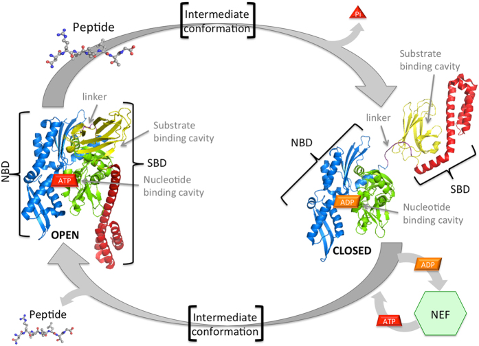
Figure 1. Hsp70 conformational cycle.
Open (ATP-bound) (PDB id: 4B9Q) and Closed (ADP-bound) (PDB id: 2KHO) end-points X-ray structure are displayed. NBD lobe I is green, lobe II is blue, linker is violet, βSBD is represented in yellow, and αSBD helices in red. Peptide-substrate bound to the SBD binding cavity of the open conformation induces the ATP hydrolysis and a large structural rearrangement to the closed conformation. This, after nucleotide exchange mediated by Nucleotide Exchange Factors (NEF), undergoes to another conformational transition, with βSBD docked to the NBD and a reduced the affinity for the peptide-substrate.

