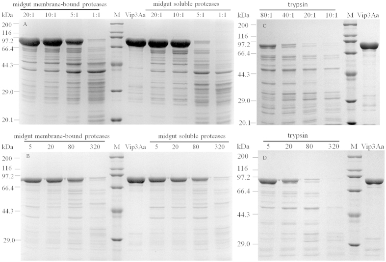
Figure 4. Proteolysis processing of Vip3Aa toxin by S. litura midgut proteases and commercial trypsin.
(A,C) Digestion of Vip3Aa toxin by different concentrations of proteases. Vip3Aa/proteases (midgut soluble, membrane-bound proteases or trypsin) ratio was showed in the figure (μg/μg). The toxins digestion with different incubation times were conducted for 5, 20, 80 and 320 min at the optimal activation concentration (B,D). Molecular mass markers (M) in kDa are indicated in the middle of the figure.

