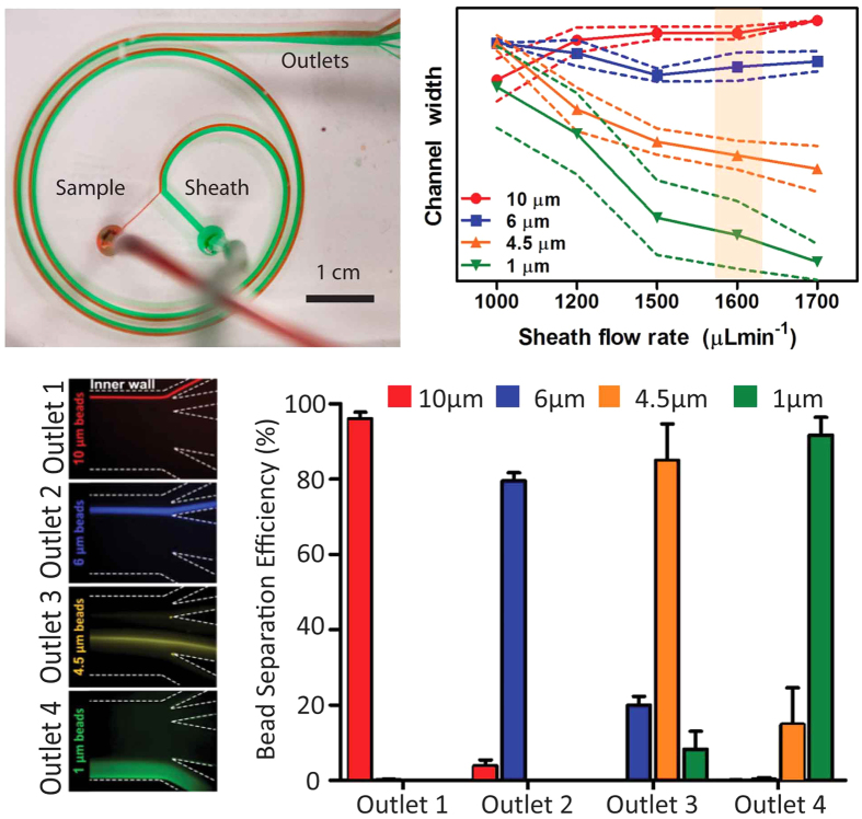
Figure 2.
(a) Fabricated PDMS spiral microchannel device showing sample (red) and sheath (green) flows. (b) Locations of streams of beads of different diameters inside the device. Solid line indicates position of centre of stream while dotted lines indicate its edges. An optimized flow rate from the highlighted range was chosen for further experiments. (c) Focused bead streams flow in separate device outlets designed to capture them. (d) Bead separation efficiency as measured using flow cytometry.

