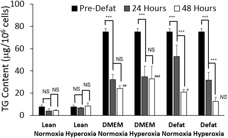
Figure 2.
Effect of defatting condition and duration on remaining triglyceride content in steatotic HepG2s. HepG2s were made steatotic by pre-incubation with free fatty acids for two days. Then, the cells were switched to basal medium with no fatty acids (DMEM), or DMEM + defatting cocktail. In each case, defatting was performed under normoxic (21% O2 v/v) or hyperoxic (90% O2 v/v). Lean controls consisted of HepG2 cells that were never made steatotic. Data shown represent the amount of triglyceride normalized to cell number in each well, measured before defatting (pre-defat), after 24 h, and after 48 h of defatting. Values are expressed as averages ± S.E.M. for n = 3 replicates. ***: p < 0.001. NS: not significantly different. Comparisons of defatted vs. lean groups: ### p < 0.001; ## p < 0.01; # p < 0.05; NS: not significantly different.

