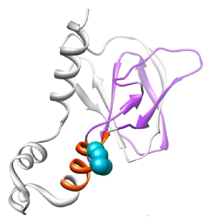
Figure 2.

Structural visualization of VHL protein with PDB ID: 1LM8 shown in ribbon representation. The protein is colored in orange [157–166: interaction with Elongin BC complex] and violet [100–155: involved in binding to CCT complex] color; the side chain of the native residue is colored cyan and shown as small balls.
