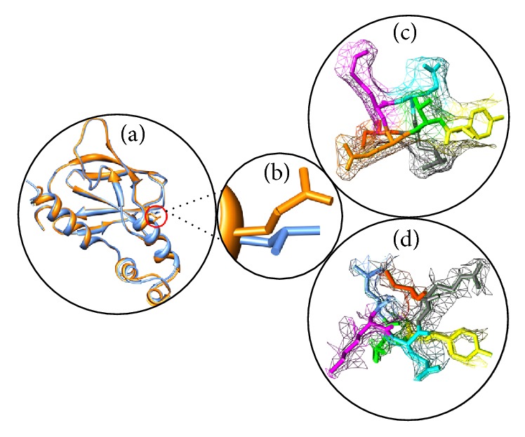
Figure 4.

(a) Superimposed structure of native and mutant structure of VHL. (b) Close-up view of native amino acid leucine (orange) and mutant amino acid glutamine (blue) in dot. (c) Local environment change visualized for native amino acid [leucine] in dot model. (d) Local environment change visualized for mutant amino acid [glutamine] in dot model. These figures were drawn using Chimera.
