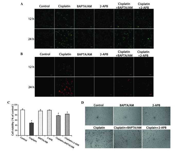
Figure 2.
Inhibition of calcium signaling decreases the level of free Ca2+ in the cytosol and mitochondria, and inhibits cell growth. (A) HeLa cells were treated with cisplatin (5 µg/ml) with or without BAPTA/AM (2.5 µM) and 2-APB (100 µM) for 12 and 24 h. The cells were incubated with the fluorescent calcium indicator, Fluo-4/AM. Calcium concentrations in the cytosol were observed by confocal microscopy (scale bar, 40 µm). (B) HeLa cells were treated with cisplatin (5 µg/ml) with or without BAPTA/AM (2.5 µM) and 2-APB (100 µM) for 12 and 24 h, and incubated with the fluorescent calcium indicator, Rhod-2. Calcium concentrations in the mitochondria were observed by confocal microscopy (scale bar, 30 µm). (C) HeLa cells were treated with cisplatin (5 µg/ml) with or without BAPTA/AM (2.5 µM) and 2-APB (100 µM) for 24 h. Cell viability was determined using the 3-(4,5-dimetrylthiazol-2-yl)-2,5-diphenyltetrazolium bromide assay. Data are presented as the mean ± standard deviation (n=3). *P<0.05 vs. control; #P<0.05 vs. cisplatin. (D) HeLa cells were treated with cisplatin (5 µg/ml) with or without BAPTA/AM (2.5 µM) and 2-APB (100 µM) for 24 h. Cell morphology was observed using an inverted phase contrast microscope at x100 magnification. BAPTA/AM, bis-(o-aminophenoxy)ethane-N,N,N',N'-tetra-acetic acid acetoxymethyl ester; 2-APB, 2-aminoethyl diphenylborinate.

