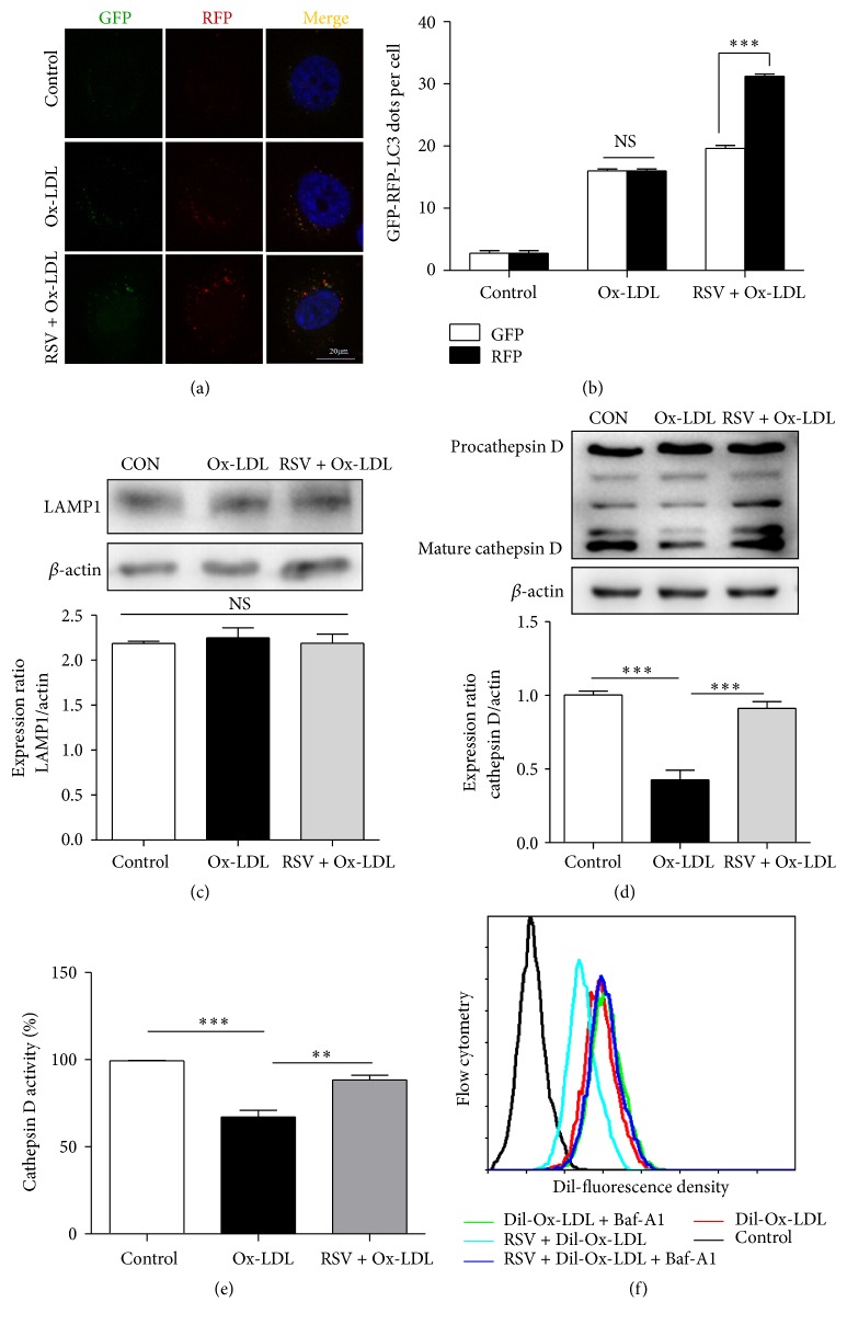
Figure 4.
Resveratrol restored lysosomal dysfunction caused by Ox-LDL in HUVECs. (a) HUVECs were transfected with RFP-GFP-tandem fluorescent LC3 cDNA for 24 h, followed by resveratrol treatment for 1 h, and subsequently incubated with Ox-LDL for another 12 h. (b) LC3 dots were determined by fluorescent confocal microscopy and quantified. At least 30 cells per group were included for the counting of RFP- and GFP-LC3 puncta/cell. Scale bar: 20 μm; ∗∗∗ p < 0.001 versus GFP; NS: not significant. Western blotting analysis of the protein level of LAMP1 (c) and cathepsin D (d) after HUVEC pretreatment with 50 μM resveratrol for 1 h, and subsequently incubation with Ox-LDL (60 μg/mL) for 12 h. Data are expressed as mean ± SEM; n = 3; ∗∗∗ p < 0.001 versus corresponding controls; NS: not significant. (e) Cathepsin D activity in HUVECs was evaluated by a fluorometric cathepsin D activity assay kit. Data are expressed as mean ± SEM; n = 3; ∗∗ p < 0.01; ∗∗∗ p < 0.001. (f) Flow cytometry detected the fluorescence density of Dil-Ox-LDL after HUVECs were treated with Dil-Ox-LDL and RSV in the presence or absence of Baf A1.

