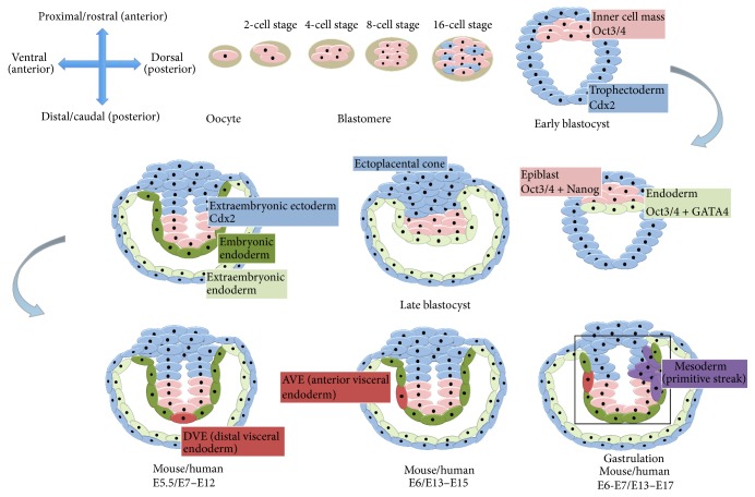
Figure 1.
Developmental stages of mouse embryo. First row (left to right), from the secondary oocyte the blastomere develops (2-cell, 4-cell, 8-cell, and 16-cell stages) to give rise to the early blastocyst formed of trophectoderm (cells that express Cdx2) and inner cell mass cells (that express Oct3/4). Later, the inner cell mass gives rise to the epiblast (cells that express Oct3/4 and Nanog) and endoderm (expressing Oct3/4 and GATA4). Second row (right to left), in the late mouse blastocyst Cdx2 positive cells give rise to the extraembryonic ectoderm and ectoplacental cone. At the same time the endoderm divides into an embryonic endoderm and an extraembryonic endoderm. The epiblast and the extraembryonic ectoderm form a cavity lined by embryonic endoderm. From the embryonic endoderm the distal visceral endoderm is formed (DVE). Third row (left to right), the DVE migrates proximally and will be known as the anterior visceral endoderm (AVE). The final image (third row, right) shows the development of the primitive streak (mesodermal cells) at the opposite (posterior) pole from the AVE. N.B. There are 2 different types of endoderm called extraembryonic and embryonic; these differ in their potency and give rise to distinct cellular derivatives. All timelines are given for mouse and human embryonic development.

