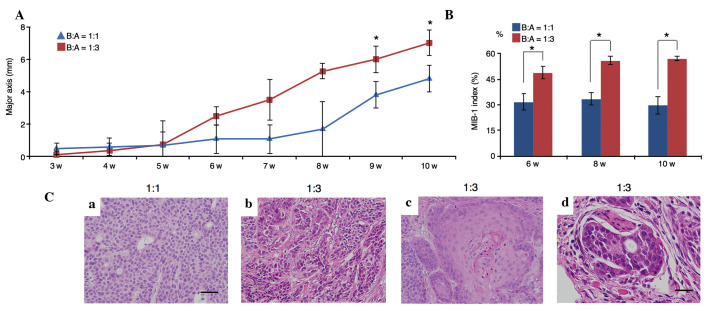
Figure 5.
Comparison of tumor growth speed, and estimation of the MIB-1 indices and histological features. (A) Tumor growth was assessed using the major axis of tumor diameter (mm). In a xenograft model in severe combined immunodeficiency mice, tumor growth of fibroblast-rich co-cultures was faster than that achieved by fibroblast-poor co-cultures, particularly at 9 and 10 weeks (*P<0.01). (B) The MIB-1 indices at 6, 8 and 10 weeks using B:A=1:3 co-cultures were significantly higher than those of the B:A=1:1 co-cultures (*P<0.01). (C) Histological features of tumors from xenografted mice [hematoxylin and eosin staining; B:A cell ratio indicated above images; (a-c) scale bar, 50 µm; (d) scale bar, 25 µm]: (a) BxPC-3 cells from fibroblast-poor co-cultures had small round nuclei; (b) in fibroblast-rich co-cultures, tumor cells exhibited pleomorphism; (c) BxPC-3 cells with rich fibroblasts exhibited differentiation to squamous cell carcinoma; (d) neural invasion was observed only in tumors initiated by fibroblast-rich co-cultures. B:A, BxPC-3:ASF-4-1. w, weeks.

