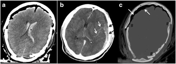
Fig. 1.

The soft-tissue-window of a cerebral CT of a young patient demonstrates the advantages of pmCT in terms of clearly showing intraventricular hemorrhage (a,*), subarachnoidal bleeding (see white arrows) along with subdural hematoma (see black arrows) on the left side (see (b) without changing the pressure-in situ. The display of CT in the bone window additionally demonstrates the multiple fragment fracture (#) of the calvarium as well as massive free intracranial air (see white arrows) (c)
