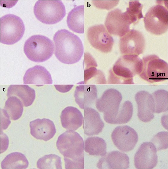
Fig. 1.

Images of erythrocytes infected with Babesia sp. in Giemsa-stained smears. a Ring form of Babesia sp. was observed in the patient’s thin blood smears prepared on July 25, 2015; b tetrads were observed in a smear of cultured blood on July 25, 2015; c, d a ring form was observed in the bone marrow smear prepared in 2005 (magnification: 100 × 10)
