Fig. 3.

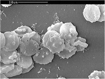
Images of erythrocytes infected with Babesia sp. obtained using an scanning electron microscope. Oval red blood cells with knobs, knob protrusion, or hollowness in the cellular membrane

Images of erythrocytes infected with Babesia sp. obtained using an scanning electron microscope. Oval red blood cells with knobs, knob protrusion, or hollowness in the cellular membrane