
Fig. 2.
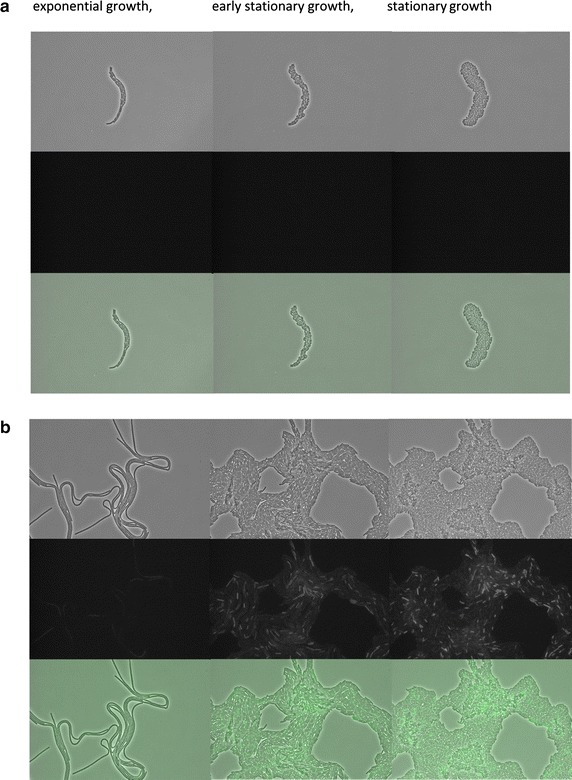
Heterogeneity of protein secretion stress in amyM-expressing cells. Time-lapse microscopy was performed of B. subtilis 168 Hm C5 (a) and 168 Hm C5 carrying plasmid pDAM coding for the secretory α-amylase AmyM (b). Strains were grown on 1.5 % agarose slides containing 25 % LB broth. Microscopy images are shown from cultures in the exponential, early stationary and stationary growth stages