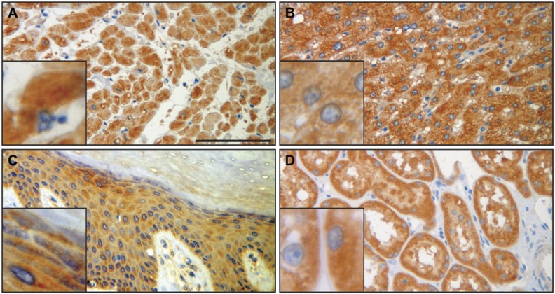
Figure 5.
SERPINB12 immunohistochemistry of heart (A), liver (B), skin epidermis (C), and kidney (D) shows diffuse, variably intense granular cytoplasmic staining in the cells of all four tissue types. Insets highlight the cytoplasmic staining. Cardiomyocytes (A) and hepatocytes (B) demonstrated the most intense staining. Scale bar = 100 µm. Images were captured on an Olympus BH2 microscope mounted with a Jenoptik ProgRes C5 camera. Images were acquired using ProgRes CapturePro 2.7 software.

