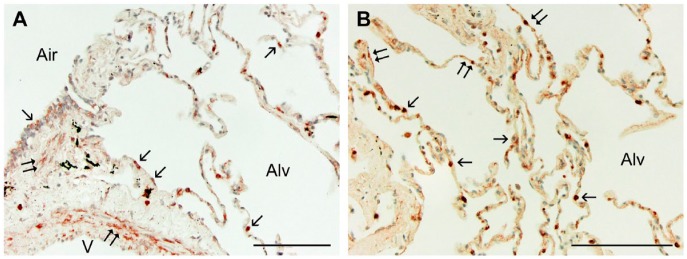
Figure 1.
Sections randomly selected from each of the three groups (10 from each, 30 total) were immunostained for Wnt5A, SP-C, or α-SMA, with appropriate controls, as described. (A) Normal human lung tissue treated for the localization of Wnt5A by immunohistochemistry showed reactivity in the smooth muscle (double arrows) of airways (Air) and vessels (V), and in the epithelium (single arrows) of airways (Air) and alveoli (Alv). (B) Higher magnification of lung parenchyma shows reactivity in some but not all cuboidal (alveolar type II; single arrows) and more squamous (alveolar type I; double arrows) cells. All sections were counterstained with methylene blue. Scale, 100 µm.

