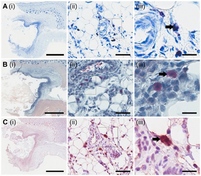
Figure 4.
Characterization and localization of mast cells. Mast cells were chemically characterized via staining with toluidine blue (A), Leder stain (B) and immunohistological methods to probe for the presence of mast cell tryptase (C). Small boxes marked in the images in the first column are sequentially enlarged in the middle and right columns. Black arrows point to punctate violet staining for mast cells (A(iii)), Leder stain positive cells (B(iii)), and mast cell tryptase positive cells (C(iii)). Scale (i) 3 mm; (ii) 60 µm; (iii) 20 µm.

