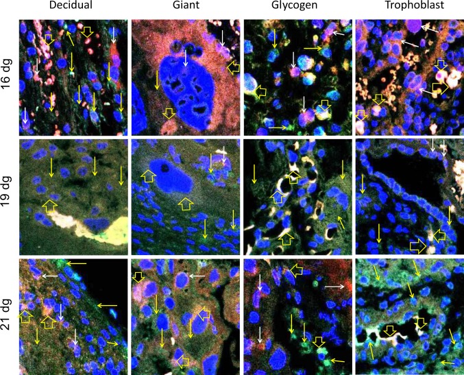
Figure 9.
Merged confocal images indicating colocalization of estrogen receptor (ER) β (red, TRITC) and metastasis-associated protein 1 (MTA1; green, FITC) in placental cells. The nuclei are labeled with 4′,6-diamidino-2-phenylindole (DAPI; blue). Expression of ERβ (red staining indicated by white arrows) and MTA1 (green staining indicated by yellow arrows) is shown. Yellow to white labeling is due to colocalization of the proteins (mixture of green and red to variable degrees due to merging of images) and is indicated by open yellow arrows. These photomicrographs show that ERβ and MTA1 are coexpressed in the cytoplasm and nucleus of decidual, glycogen, giant, and trophoblast cells on 16 dg, 19 dg, and 21 dg. Original magnification, 400×. (The color version of this article is available at http://rs.sagepub.com.)

