Fig. 3.
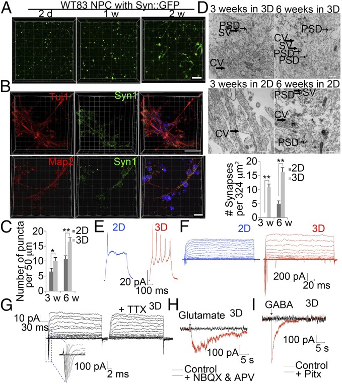
Rapid maturation of human iPSC-derived neurons in 3D hydrogels. (A) Representative images of human iPSC-derived NPCs infected with Syn::GFP lentivirus after neuronal differentiation for indicated times in 3D hydrogels. (Scale bar, 200 μm.) (B) Representative images of human iPSC-derived neurons stained for Tuj1 or Map2 (red), Syn1 (green), and DAPI (blue) in 3D hydrogel. (Scale bar, 60 μm.) (C) Quantification of Syn1 puncta on Map2 neurites. Bars represent means of 50 neurons per condition; *P < 0.05, and **P < 0.01; n = 3. (D) Representative images of ultrastructural investigation of synaptogenesis by transmission electron microscopy in control NPCs differentiated in 3D or 2D system at indicated times. CV, large clear vesicle; SV, synaptic vesicle. Quantification of numbers of synapses in control NPCs differentiated in 3D or 2D system at indicated times. Bars represent means; **P < 0.01 by; n = 3. (Scale bar, 500 nm.) (E) Representative whole-cell current clamp recordings of human iPSC-derived neurons after 3 wk differentiation in 3D or 2D system. Spiking activity was examined following current injection. (F) Representative voltage clamp recording of a 3D or 2D cultured neuron held at −75 mV and then stepped to a series of voltages (−80 to +20 mV) in 5-mV increments. (G) Representative voltage clamp recording of a 3D cultured neuron before and after application of TTX. TTX, 1 μM tetrodotoxin. (H) Response to glutamate in the presence and absence of the glutamate receptor blockers 20 μM NBQX and 50 μM APV. (I) Response to GABA in the presence and absence of 50 μM picrotoxin (Pitx).

