Fig. 3.
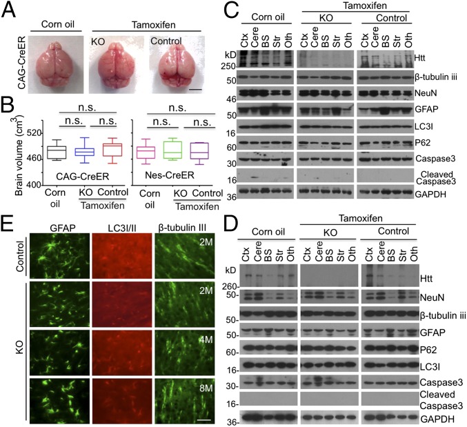
Normal brains in adult mice that have depleted Htt. (A) The brain photos of 2-mo-old ubiquitous KO mice that had been injected with tamoxifen for 5 d. (B) Brain volumes of 2-mo-old ubiquitous KO mice in A and neuronal KO mice that were injected with tamoxifen at 2 mo of age and used to isolate their brains at the 90th day after tamoxifen injection (one-way ANOVA test, ubiquitous KO: n = 7, P = 0.8759, F = 0.1335; n = 7; neuronal KO: P = 0.9665, F = 0.03416; n.s. represents no significant differences). (Scale bar, 3 mm.) (C and D) Western blotting analysis of the brain tissues from 2-mo-old ubiquitous (C) and neuronal (D) KO mice. Htt, neuronal (NeuN, β-tubulin III), astrocytic (GFAP), autophagic (P62, LC3I/II), apoptotic (cleaved caspase3), and GAPDH were detected by their antibodies. Control is heterozygous floxed Htt/CAG-CreER (C) or heterozygous floxed Htt/Nestin-CreER (D) mice injected with tamoxifen and homozygous floxed Htt/CAG-CreER (C) or homozygous floxed Htt/Nestin-CreER (D) mice injected with corn oil. KO is homozygous floxed Htt/CAG-CreER (C) or homozygous floxed Htt/Nestin-CreER (D) mice injected with tamoxifen. See Fig. 1 for abbreviations key. (E) Immunostaining of GFAP, LC3I/II, and β-tubulin III in the cortex of 2-, 4- and 8-mo-old ubiquitous KO and control mice. (Scale bar, 50 μm.)

