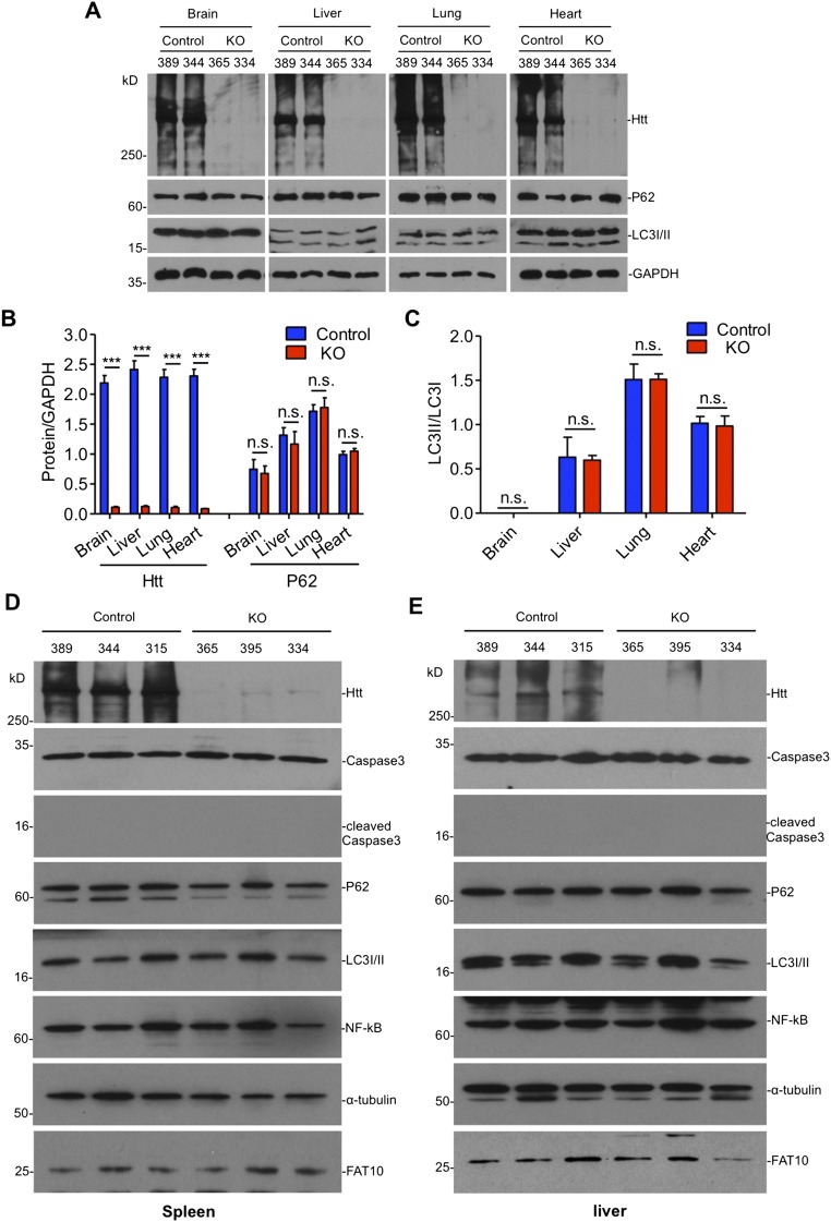
Fig. S1.
The expression of autophagy markers in the brain and peripheral tissues of adult ubiquitous Htt KO mice. (A–C) Representative immunoblots (A) and quantification of (B) Htt, P62, and (C) LC3I/II ratio in ubiquitous KO and control mice (n = 3 independent experiments). (D and E) Western blotting of the spleen (D) and liver (E) in control and ubiquitous KO mice. The blots were probed by antibodies to cleaved caspase3, P62, LC3I/II, NF-κB, and FAT10 after tamoxifen injection for 3 d. Alpha-tubulin was probed as a loading control. Animal ID numbers are indicated. Ctl: heterozygous floxed Htt/CAG-CreER mice injected with tamoxifen; KO: homozygous floxed Htt/CAG-CreER mice injected with tamoxifen.

