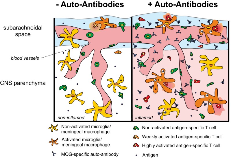
Fig. 6.
Schematic depiction summarizing how MOG AAbs enhance autoimmune CNS disease. (Left) In the absence of AAbs, autoantigen-specific effector T cells scan the endothelial cells of the BBB. Few cells penetrate the BBB and are suboptimally reactivated by local APCs (microglia/meningeal macrophages). The reactivated T cells initiate a cascade of inflammatory events, which might eventually lead to autoimmune disease development. (Right) When AAbs are present in the blood circulation, they pass through the BBB along with the autoantigen-specific T cells and locally bind to their specific antigen. The antibody complexes are taken up by resident macrophages/microglia that then efficiently present the myelin antigen to the autoantigen-specific effector T cells. The enhanced reactivation of the effector T cells leads to strongly increased inflammation of the CNS parenchyma and accelerates and aggravates the clinical disease.

