Fig. 5.
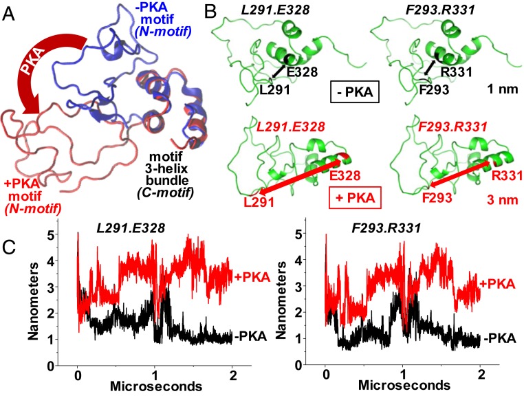
(A) Microsecond-long MD simulations were performed to determine the effect of phosphorylation on the MyBP-C motif (residues 259–353). The accessibility of the three α-helix bundles changes as the disordered region of the motif rotates away from the helices (blue to red conformation). (B) Distance measurements were simulated between three-helix bundle residues (E328 or R331) and disordered region residues (L291 or F293) with (red) and without (black) PKA phosphorylation. (C) Trajectories of the distances between these pairs with or without phosphorylation.

