Fig. 1.
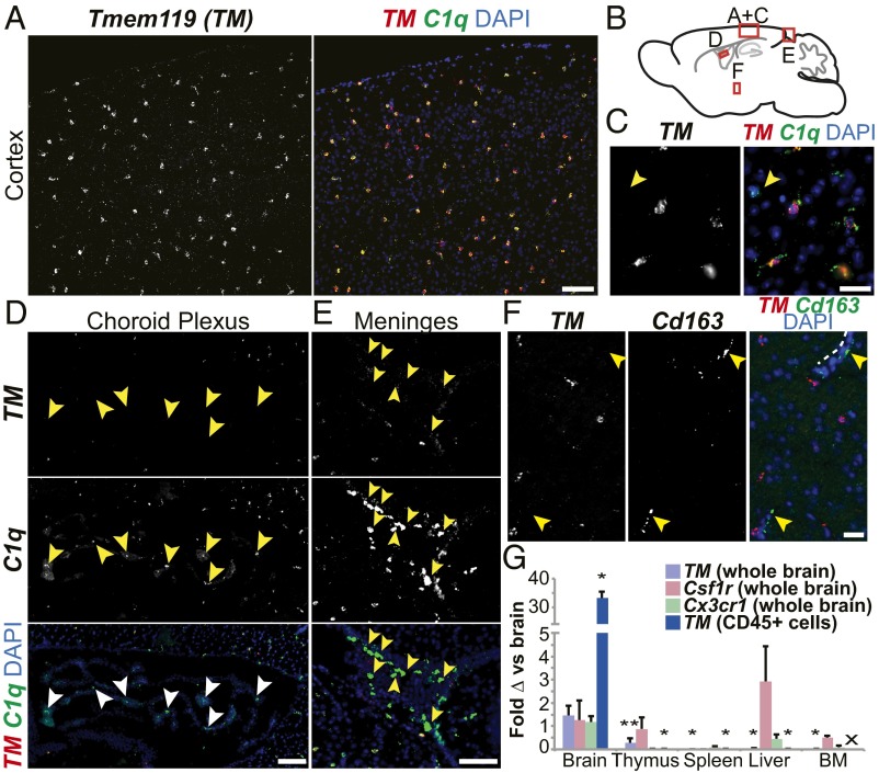
Tmem119 is specifically expressed by parenchymal myeloid cells in the CNS. (A) In situ hybridization of mouse brain revealed widespread Tmem119 expression by myeloid (C1q+) cells. Composited from two adjacent imaging fields. (B) Sagittal brain schematic of panel locations. (C) Higher power showing C1q+ Tmem119+ and a rare parenchymal C1q+Tmem119− cell (arrow). C1q+ cells in the choroid plexus (D) and meninges (E) were Tmem119− (arrows). (F) Tmem119 mRNA was not detected in Cd163+ perivascular macrophages (arrows; dotted line highlights one vessel). (G) qPCR analysis of Tmem119, Csf1r, and Cx3cr1 expression by whole tissues and Tmem119 by CD45+ cells compared with average expression of each gene in whole brain. Data presented as mean ddCT ± SEM; *P < 0.01 compared with whole brain Tmem119; **P < 0.02 by ANOVA with post hoc Tukey HSD for dCT values. n = 4–9 except BM, CD45+ liver, CD45+ thymus where n = 2. x, no sample for CD45+ BM. (Scale bars: 100 μm in A, D, and E; 40 μm in C and F.) See also SI Appendix, Fig. S1.

