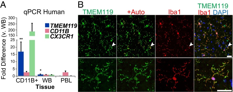
Fig. 6.
Tmem119 is also highly enriched in and specific to human microglia. (A) Relative expression of TMEM119, CD11B, and CX3CR1 mRNA in human peripheral blood leukocytes (PBL), whole brain (WB), and CD11B+ brain cells *P < 0.01 by ANOVA with Tukey HSD. Error bars show SEM, for n = 2 independent samples per tissue type. (B) Representative confocal images of 51-y-old normal appearing cortical tissue showing localization of TMEM119 (green) to highly ramified, Iba1+ cells (red) at low (Upper) and high (Lower) magnification. Autofluorescence channel (red) overlays green to highlight nonspecific fluorescence from lipofuscin (arrows), which are absent in Lower because of Sudan Black treatment. (Scale bars, 30 μm.)

