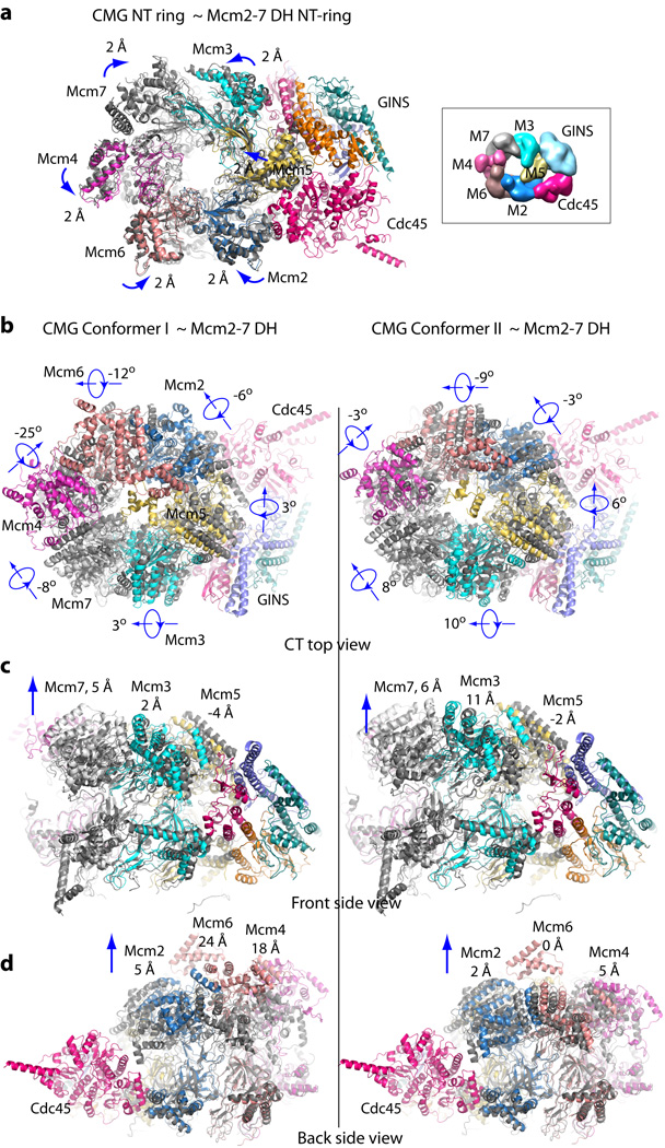
Figure 5. Remodeling changes between the Mcm2-7 in the double-hexamer (DH) and in the active CMG conformers.
(a) Superimposition of the Mcm2-7 NTD-tier ring of CMG (color cartoon representation) with that of the inactive double-hexamer (gray cartoon representation). The double-hexamer Mcm2-7 structure is (EMDB 6338)13. The direction and distance of rigid body domain movements are noted by each subunit. At the right (boxed) is a surface representation of the NTD view of the previously reported 3D density map of CMG (EMDB 6463)16. (b) Comparison of the top view of the CTD-tier of Mcm2-7 of the double-hexamer with the Mcm2-7 CTD-tier of CMG conformer I (left column) and CMG conformer II (right column), and in: (c) Front side view, and in (d) Back side view. Each CTD AAA+ domain undergoes a combination of rotation and translation, with the degree of rotation shown in (b) and the vertical translation in (c) for Mcm3, Mcm5, Mcm7, and (d) for Mcm2, Mcm4, and Mcm6. Another significant change is the insertion of the Mcm5 WHD into the interior channel in the active helicase, but this is not illustrated here because the deposited PDB coordinates of the inactive Mcm2-7 hexamer do not contain the Mcm5 WHD. The Mcm5 WHD in DH is documented and explained in Extended Data Figure 3d–e13.

