
Figure 2.
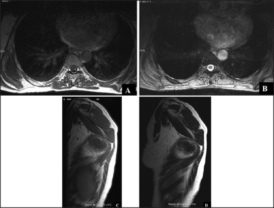
(a and b) MRI image showing focal ill-defined altered signal intensity lesion in the right posterolateral chest wall deep to serratus anterior muscle. The lesion is isointense to muscle in T1 and T2WI images (axial) (c and d) T1 and T2WI images (sagittal) of the lesion