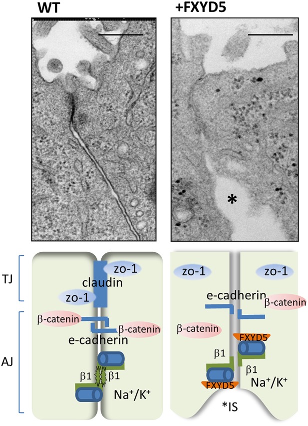
Figure 1.

FXYD5 impairs cell–cell junction formation. (Upper) High magnification of electron microscopy images of normal and FXYD5 transfected M1 cell monolayer. Large expansions of interstitial spaces (IS) located just under the TJ evoked by FXYD5 expression (asterisks). Bar = 200 nm. The EM images were originally published in (Lubarski et al., 2011). (Lower) Experimental mode of FXYD5 function. FXYD5 expression in M1 polarized monolayer results in down regulation in membrane expression of TJ markers ZO-1 and occludin. E-cadherin membrane localization remained intact, while β-catenin, has been redistributed and appeared perpendicular to the membrane plane. A reduced form of β glycosylation has been observed, indicating an additional condition for promotion of “leaky” epithelia.
