
Figure 11 (A and B).
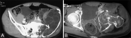
CT images of bone hydatid. (A) There is expansion of left iliac bone with destruction of anteromedial cortex by a multiloculated cystic mass. Transarticular extension to sacrum is also seen (B) Involvement of ischium, pubis, hip joint, and proximal femur is evident with extension into the adjacent muscles