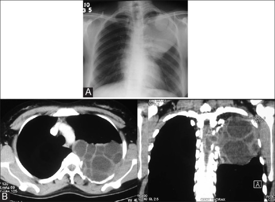
Figure 12 (A and B).

Costal echinococcosis with intraspinal extension in a 22-year-old female who presented with numbness of left lower limb. (A) Chest radiograph shows a homogenous opacity in the upper part of left hemithorax. A round, well-defined opacity is seen in its upper part. Erosion of posterior parts of fourth and fifth ribs is seen (B) CT images in the axial and coronal plane reveal a multiloculated cystic mass in the posterior left hemithorax with erosion of adjacent rib and extension into the spinal canal
