
Figure 13.
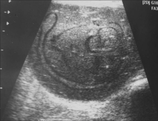
Ultrasound image of a 22-year-old female shows a type IV hydatid cyst in the thigh muscle. A well-defined hypoechoic mass with posterior enhancement and separated membranes within is seen
Official websites use .gov
A
.gov website belongs to an official
government organization in the United States.
Secure .gov websites use HTTPS
A lock (
) or https:// means you've safely
connected to the .gov website. Share sensitive
information only on official, secure websites.
Ultrasound image of a 22-year-old female shows a type IV hydatid cyst in the thigh muscle. A well-defined hypoechoic mass with posterior enhancement and separated membranes within is seen